St17-8.45hpf
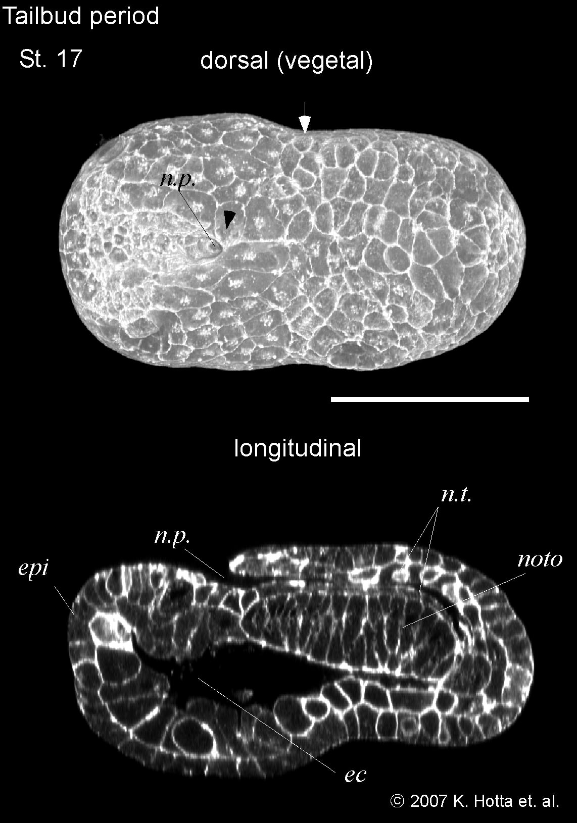
initial tailbud I (8.45 hours), there is the first indication of a separation between tail and trunk territories. The tail is not bent and has the same length as the trunk. The neural tube closure in the posterior territory finished and the neuropore move more anterior. The notochord cells are arranged in two inter-digitating rows but the notochord cells have not finished intercalation.
Three-dimensional images (upper) and cross-section images (middle). Anterior is left in all panels. Arrow indicates the position of pigment cells. Arrowheads indicate the cilia of epidermal sensory neurons. b.p., blastopore; n.p., neuropore; epi, epidermis; ec, endodermal cavity; en, endoderm; es, endodermal strand; b, brain; mu, muscle; g, germ line cells; noto, notochord; n.t., neural tube; ec, endodermal cavity; t.epi.m-l.r, tail epidermis medio-lateral row; t.epi.m.r, tail epidermis medial row; t.epi.l.r, tail epidermis lateral row; va, vacuole; p, palps. In I, embryo direction is shown by A: anterior, P: posterior, D: dorsal, V: ventral. Scale bar = 50 micro m.