St26-17.5hpf
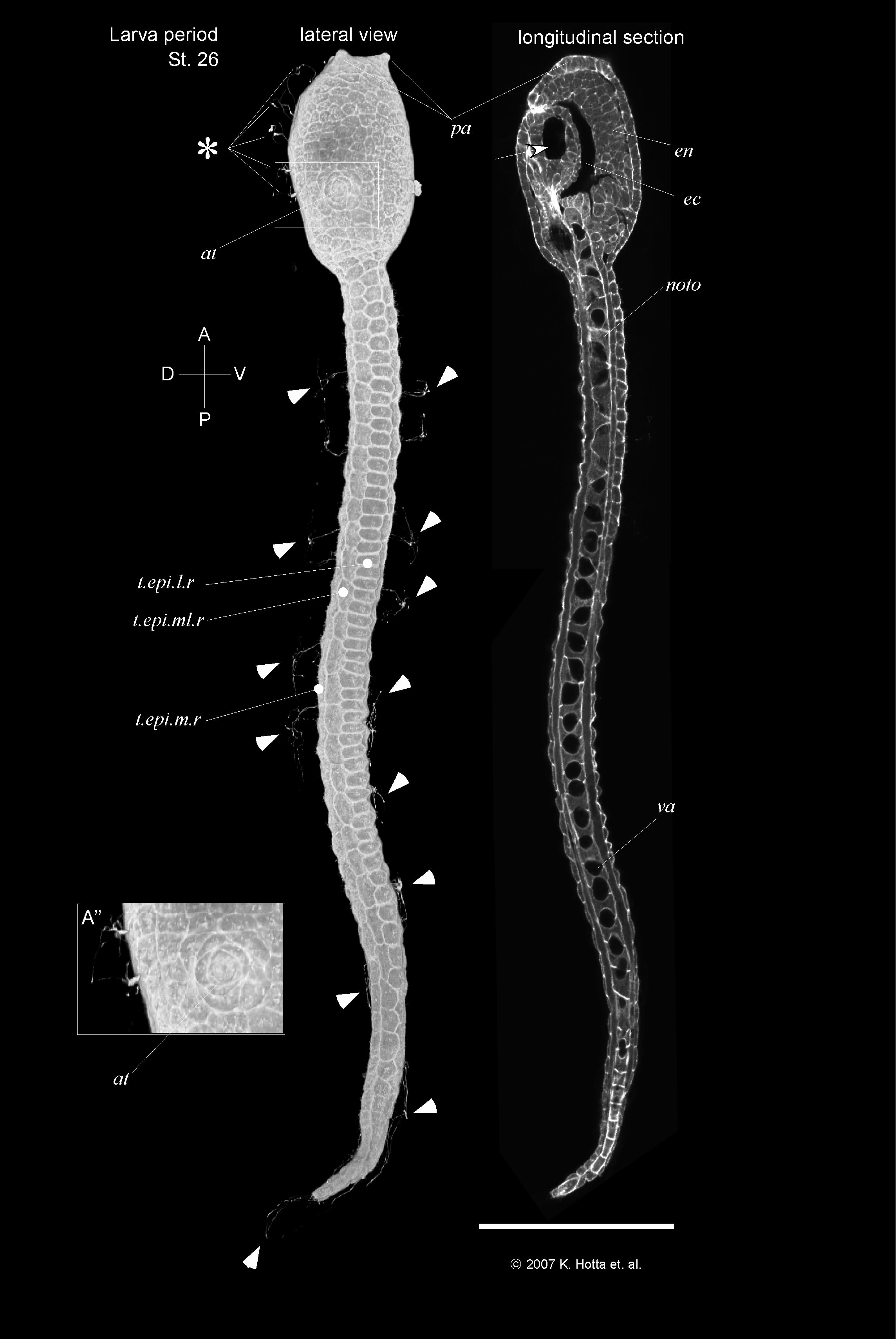
hatching larva (17.5 hours), the head adopts an elongated rectangular shape. Cilia of caudal epidermal sensory neuron are growing. These cilia project into the fin tunic. Such structures are found also in the head (Hotta, unpublished data). A pair of atrial primordia is well observed.
Arrowheads indicate cilia from tail epidermal sensory neurons. Arrow indicates the position of pigment cells. Asterisk indicates cilia from epidermal sensory neuron in head. n.p., neuropore; epi, epidermis; en, endoderm; es, endodermal strand; b, brain; mu, muscle; g, germ line cells; noto, notochord, n.t., neural tube; ec, endodermal cavity; t.epi.m-l.r, tail epidermis medio-lateral row; t.epi.m.r, tail epidermis medial row; t.epi.l.r, tail epidermis lateral row; va, vacuole; pa, palps; at, atrial primordia. A: anterior, P: posterior, D: dorsal, V: ventral. Scale bar = 50 micro m.