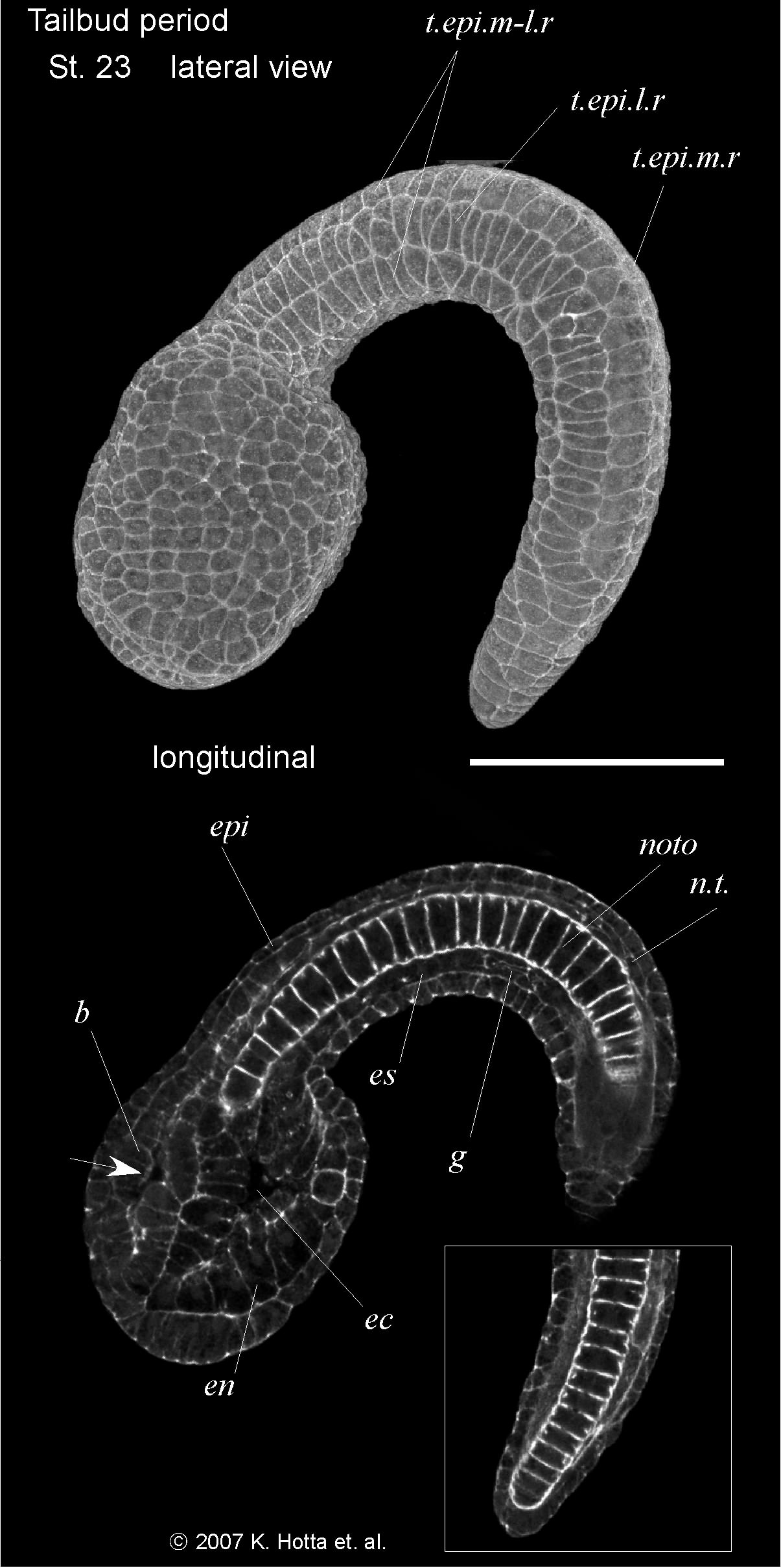
St23-11.9hpf
late tailbud I (11.9 hours), the initiation of the pigmentation of the otolith under a dissecting microscope can be observed. The tail is acutely curved with the tip close to the anterior end of the trunk.
Three-dimensional images (upper) and cross-section images (lower). Anterior is left in all panels. Arrow indicates the position of pigment cells. Arrowheads indicate the cilia of epidermal sensory neurons. b.p., blastopore; n.p., neuropore; epi, epidermis; ec, endodermal cavity; en, endoderm; es, endodermal strand; b, brain; mu, muscle; g, germ line cells; noto, notochord; n.t., neural tube; ec, endodermal cavity; t.epi.m-l.r, tail epidermis medio-lateral row; t.epi.m.r, tail epidermis medial row; t.epi.l.r, tail epidermis lateral row; va, vacuole; p, palps. In I, embryo direction is shown by A: anterior, P: posterior, D: dorsal, V: ventral. Scale bar = 50 micro m.